Transcript
- [Narrator] This video is for demonstration
and educational purposes only.
Please follow your own institutional guidelines
on sterility and safety.
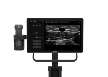
A linear array transducer with a venous exam type
is used to perform an ultrasound-guided insertion
of a peripherally inserted central catheter
via a transverse approach.
The patient is placed in a supine position
with the arm extended 90 degrees at the patient's side
and externally rotated.
The transducer is placed transversely
in the medial upper arm, superior to the antecubital fossa
and inferior to the axilla.
The orientation marker should be directed
to the patient's right.
The basilic vein is seen
as an anechoic circular compressible structure
that is superficial and medial
to the brachial vein and artery.
Deeper within the image,
the hyperechoic humerus can be seen.
Adjust the transducer
so the basilic vein is centered within the ultrasound image.
With the needle positioned just superior to the transducer,
slowly insert the needle
next to the center line on the transducer.
The needle will appear as a small bright dot
in the ultrasound image.
When the needle tip appears,
slide the transducer a short distance away
until the tip is no longer visible.
Once again, advance the needle slowly
until it is visible in the ultrasound image.
Repeat the process to ensure
that the needle tip is advanced
under direct ultrasound guidance.
Continue advancing the needle
until the tip is seen to indent
and then puncture the basilic vein.
Confirm that the needle tip lies
in the mid portion of the basilic vein lumen
by rotating the probe 90 degrees
to get an in plane longitudinal view of the needle
within the vein.
This technique can also be used to place the PICC line
within the deep brachial or cephalic veins.
How to: Ultrasound Guided PICC Line
A linear array transduer with a venous exam type is used perform an ultrasound guided insertion of a peripherally inserted central catheter via a transverse approach.