Transcript
- [Narrator] This video is for demonstration
and educational purposes only.
Please follow your own institutional guidelines
on sterility and safety.
A linear ray transducer
with a venous or PIV exam type is used to perform
an ultrasound-guided peripheral IV device insertion
via a transverse approach.
The patient is placed in a supine or semi-upright position
with the examiner positioned on the same side
that the IV device will be inserted.
The ultrasound machine should be positioned
to provide an ergonomic direct line of sight.
The transducer is placed on the distal forearm
just above the wrist region
with the orientation marker directed
towards the clinician's left shoulder.
A vascular assessment of the forearm
or another intended insertion site is performed
to identify a vein with suitable size and location,
away from the areas of flexion or motion.
The ultrasound image should be optimized
for depth and gain.
Veins are seen as hypoechoic round structures
that are compressible with slight transducer pressure.
It is important to maintain minimal transducer pressure
during the examination to avoid vessel compression.
Adjust the transducer so the intended target is centered
within the ultrasound image.
With the needle positioned just superior
to the transducer, slowly insert the needle
next to the center line on the transducer.
The needle will appear as a small, bright dot
in the ultrasound image.
When the needle tip appears,
slide the transducer a short distance away
until the tip is no longer visible.
Once again, advance the needle slowly
until it is visible in the ultrasound image.
Repeat the process to ensure
that the needle tip is advanced
under direct ultrasound guidance.
Continue advancing the needle
until the tip is seen to indent and then puncture the vein.
Confirm that the needle tip lies in the mid portion
of the vein by rotating the probe 90 degrees
to get an in-plane longitudinal view of the needle
within vessel lumen.
The catheter is then slowly advanced off the stylet
and into the vessel to complete the procedure.
This procedure can also be performed entirely
in an in-plane approach
based on the clinician's experience and preference.
How to: Ultrasound Guided PIV
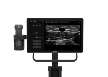
This video details how the use of ultrasound imaging can identify deep and nonpalpable veins that can accommodate the placement of an intravenous catheter. The video also depicts how Doppler color flow is used to differentiate the brachial artery from other anatomical structures.