Metacarpophalangeal (MCP) Joint Synovitis: Early Rheumatoid Arthritis (RA) (Seronegative)
- Obtaining a video clip helps with diagnosis. In this video clip, you can see some noise artifact that can occur with transducer movement. This is common.
- Also, another good practice is to raise the color Doppler gain until a noisy artifact appears under the cortical surface of the bone, then reduce the gain until this noise just barely disappears.
- This represents a “noisy threshold of gain,” which means your gain setting is high enough.
- You don’t want too much noise artifact, but you don’t want too little, which can cause you to miss data.
- Any color Doppler that is seen under the bone is considered artifact, and if shown in a still image, the color Doppler seen above the cortex should also be considered suspicious of being artifact.
- Video clips can clear up any doubts of artifact vs. true color Doppler flow, if the flow persists and stays in the frame and is above the cortical surface (but not touching) it can be considered to be truly color Doppler flow.
- Be suspicious about flow that is lining the cortical surface, this can be an artifact also.
- To confirm these findings, scan the same area in transverse.
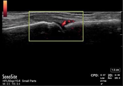
- This color Doppler image in the short axis shows the same synovial fluid with an increase in color flow.
- There are normal interdigital vessels.
- However, those that are coursing right over the base of the proximal phalanx are not normal
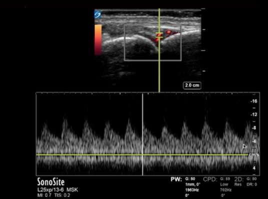
This image shows synovial fluid with pulse wave Doppler.
- Set the sample box directly over one of the vessels.
- The wave formation can show that the object is arterial or venous and not an artifact.
- This particular image shows arterial flow.
- Venous flow would be flatter with some ups and downs in its shape.
- Artefactual objects would be more scattered along the baseline.