Transcript
- [Narrator] This video is for demonstration
and educational purposes only.
Please follow your own institutional guidelines
on sterility and safety.
A linear ray transducer with a venous exam type
is used to perform an ultrasound-guided insertion
of a subclavian vein catheter via a transverse approach.
The patient is in a supine position with the head neutral.
The operator should be positioned
on the same side that the catheter will be inserted.
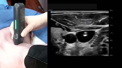
The transducer is placed transversely on the vessel,
just inferior to the midportion of the clavicle,
with the orientation marker directed to the patient's head
at a 12 o'clock position.
The subclavian vein is seen as an anechoic circle,
just inferior and deep to the clavicle.
The transducer should be slowly moved
one to two inches laterally,
with the face of the transducer staying below the clavicle
to obtain the best view of the distal subclavian
and proximal axillary vein.
It is important to note that the lung lies
directly posterior to the vessel,
so posterior wall puncture of the vein should be avoided.
Adjust the transducer so the vein is centered
within the ultrasound image.
With the needle positioned just superior to the transducer,
slowly insert the needle
next to the center line on the transducer.
The needle will appear as a small bright dot
in the ultrasound image.
When the needle tip appears,
slide the transducer a short distance medially
until the needle is no longer visible.
Once again, advance the needle slowly
until it is visible in the ultrasound image.
Repeat the process to ensure that the needle tip
is advanced under direct ultrasound guidance.
Continue advancing the needle until the tip
is seen to indent and then puncture the vein.
Confirm that the needle tip lies
in the midportion of the vein by rotating the probe
90 degrees to get an in-plane longitudinal view
of the needle within the subclavian proximal axillary vein.
手順:超音波ガイド下鎖骨下静脈中央ラインの挿入
横方向アプローチによる鎖骨下静脈カテーテル挿入の教育用超音波デモンストレーションです。患者は仰臥位で、頭部は中立位に保たれています。鎖骨下静脈を同定するため、鎖骨の下にリニアプローブを当てます。後方肺穿刺を避けながら、リアルタイム画像のガイド下で針を挿入し、縦断像で静脈内への挿入を確認します。
関連ビデオ (1)