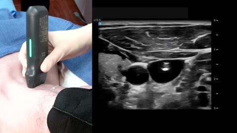
of a subclavian vein catheter via a transverse approach.
The patient is in a supine position with the head neutral.
The operator should be positioned
on the same side that the catheter will be inserted.
The transducer is placed transversely on the vessel,
just inferior to the midportion of the clavicle,
with the orientation marker directed to the patient's head
at a 12 o'clock position.
The subclavian vein is seen as an anechoic circle,
just inferior and deep to the clavicle.
The transducer should be slowly moved
one to two inches laterally,
with the face of the transducer staying below the clavicle
to obtain the best view of the distal subclavian
and proximal axillary vein.
It is important to note that the lung lies
directly posterior to the vessel,
so posterior wall puncture of the vein should be avoided.
Adjust the transducer so the vein is centered
within the ultrasound image.
With the needle positioned just superior to the transducer,
slowly insert the needle
next to the center line on the transducer.
The needle will appear as a small bright dot
in the ultrasound image.
When the needle tip appears,
slide the transducer a short distance medially
until the needle is no longer visible.
Once again, advance the needle slowly
until it is visible in the ultrasound image.
Repeat the process to ensure that the needle tip
is advanced under direct ultrasound guidance.
Continue advancing the needle until the tip
is seen to indent and then puncture the vein.
Confirm that the needle tip lies
in the midportion of the vein by rotating the probe
90 degrees to get an in-plane longitudinal view
of the needle within the subclavian proximal axillary vein.
Anleitung: Ultraschallgesteuerte Anlage eines zentralen Katheters in die Vena subclavia
Ultraschall-Demonstration zur Einleitung eines Katheters in die Vena subclavia mittels eines transversalen Zugangs. Der Patient liegt in Rückenlage mit neutraler Kopfhaltung. Eine lineare Sonde wird unterhalb des Schlüsselbeins platziert, um die Vena subclavia zu lokalisieren. Die Nadel wird unter Echtzeit-Führung vorgeschoben, wobei eine Punktion der hinteren Lunge vermieden wird, und in der Längsschnittansicht innerhalb der Vene bestätigt.