Skip to main content
Metacarpophalangeal (MCP) Joint Dorsal: Normal vs Inflammatory Synovitis
Metacarpophalangeal (MCP) Joint Dorsal: Normal vs Inflammatory Synovitis
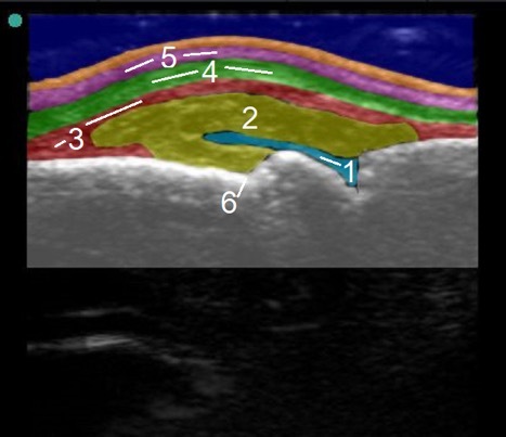
- Synovial Fluid
- Joint Capsule
- Aponeurosis
- Extensor Tendon
- Extensor Hood
- Metacarpal Notch
- Small swelling of the MCP joint is commonly seen with rheumatoid arthritis.
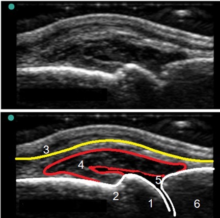
- Distal Metacarpal Head
- Metacarpal Notch
- Extensor Tendon
- Synovial Membrane
- Dorsal Recess
- Proximal Phalanx
- This image shows a severe case of inflammation.
- It’s early enough that we don’t see erosions in the base of the metacarpal notch.
- The extensor tendon surface looks bowed and swollen.
- The simple effusion (synovial fluid) is compressible.
- This case of rheumatoid arthritis has not significantly eroded the bone yet.
- It’s best to view this with color Doppler to see if there is active synovitis, which is definitely suspect in this case.