Courses

The following point-of-care ultrasound courses are recommended for you based on what we know about your interests. You can follow the suggested curriculum, or explore other materials at your own pace. To view other available courses, click the courses button at the top of this page.

TEE Hero Image

*NEW* Resuscitative TEE - Transesophageal Ultrasound
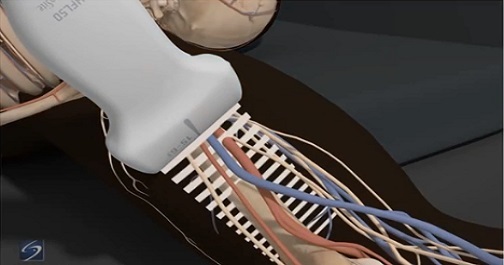
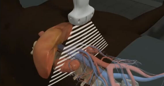
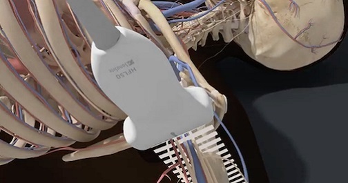
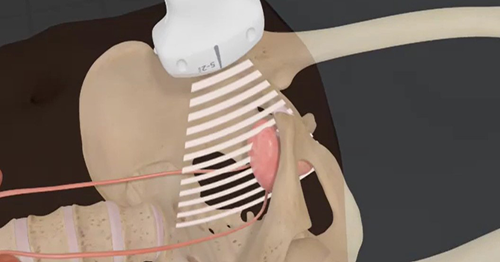
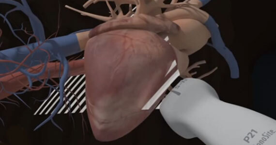
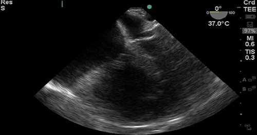
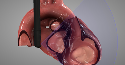
 (TEU)

Point of care Transesophageal Ultrasound (TEU) is an emerging modality that offers bedside clinicians the ability to assess critically ill patients quickly. A Transesophageal Echocardiogram, or TEE, is an alternative way to perform an echocardiogram. A specialized transducer containing an ultrasound transducer at its tip is passed into the patient's esophagus. This allows image and Doppler evaluation which can be recorded.

aorta

Aorta

After completing the aorta ultrasound course, participants should be able to: identify the anatomical structures visualized during the aorta examination, recognize the various types of abdominal aortic aneurysms and perform their associated measurements, and determine the preferred transducer to perform the aorta exam.

Axillary Nerve Block

Axillary Nerve Block

The axillary block is a plexus block at the terminal branches of the brachial plexus, designed to anesthetize the major motor and sensory nerves in the distal arm, elbow, wrist, forearm, hand, and fingers. Course participants will learn the anatomy of the axillary vessels and musculocutaneous nerve and the technique for performing an axillary nerve block.

Cardiac Imaging 1 Hero

Cardiac Imaging 1

How do you angle the transducer to get a Parasternal Long Axis (PLAX) view of the heart in the Emergency Department? Which kind of transducer should you use? How is the cardiology orientation different? And how, exactly, do you measure fluid responsiveness using the Inferior Vena Cava (IVC)?

Cardiac Imaging 1 introduces these questions (and answers!), and much more. Develop an understanding of how emergency medicine and cardiology applications differ, and test your knowledge of different views of the heart.

Doppler Principles

Cardiac Imaging 2 - Doppler Principles (English Only)

Doppler echocardiography is the language of flow in and around the heart. In order to evaluate hemodynamics in and around cardiac valves, cardiac pressures or in the calculation of Stroke Volume (SV), one must speak the language of Doppler.

This introduction to Doppler principles and how they relate to point-of-care echocardiography will frame the knowledge you need to engage some particularly valuable hemodynamic techniques!

Stroke Volume

Cardiac Imaging 2 - Stroke Volume (English Only)

Determination of Stroke Volume (SV) is perhaps the most essential of all the “advanced” techniques for point-of-care echocardiography. It’s considered advanced, because it relies on quantitative spectral Doppler techniques, which are not routinely part of the core point-of-care echocardiography curricula offered for most specialties at the time of this publication. This technique is the most practical and intuitive gateway to hemodynamic understanding of echocardiography and is a powerful adjunct in the assessment of Left Ventricular (LV) function.

Central Line Management Program

Central Line Management Program

Central Line placement is one of the most common hospital procedures, and yet it is not without risk. Ultrasound-guided Central Venous Catheter (CVC) placement reduces some of the more severe risks, such as pneumothoraces and CLABSI. Learn the procedures, techniques, and best practices associated with placing a central line under ultrasound guidance while minimizing risks of iatrogenic complications.