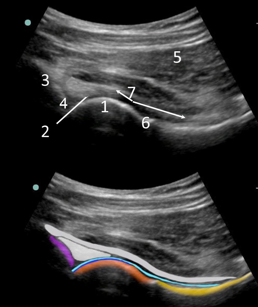
Note: The 2-3 mm anechoic or hypoechoic area above the subchondral bone is articular cartilage and not to be confused with an effusion.
Note the acetabulum (3) and labrum (4).
Superficial to the labrum is the iliofemoral ligament.
The iliofemoral ligament drapes from its combined capsular origin over the femoral head (1), where it becomes a dedicated ligament, and drapes distally over femoral neck (6), attaching at the intertrochanteric line.