EMED

Pelvic First Trimester Pregnancy

Pelvic: First Trimester Pregnancy Evaluation

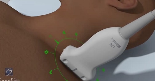
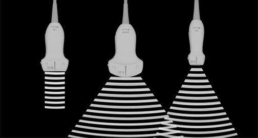
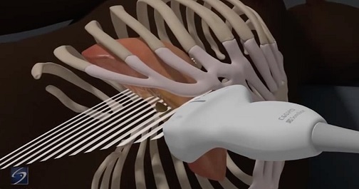
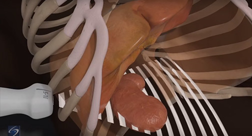
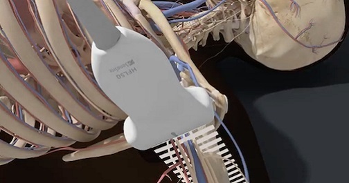
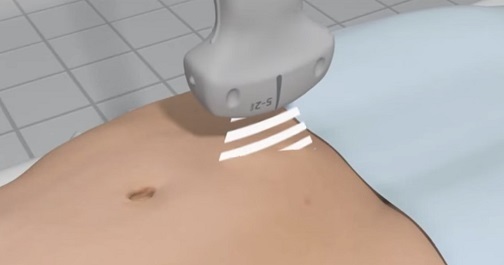
Understand the basic ultrasound views and techniques associated with performing an ultrasound pelvic examination for first trimester pregnancy, surrounding anatomical structures, and proper equipment settings. In addition, support literature, case studies, pathology images, and videos may be included for review. 

Popliteal Sciatic Nerve Block

Popliteal-Sciatic Nerve Block

Review and learn to identify anatomical structures visualized during a popliteal sciatic nerve block with ultrasound guidance; determine the locations of the popliteal artery and vein, and how to locate the perineal and tibial nerve branch, which is ideal for the nerve block placement. 

Ultrasound Guided Paracentesis

Ultrasound-Guided Paracentesis

After completing the Ultrasound-Guided Paracentesis course, viewers should be able to explain the basic outline of ultrasound-guided paracentesis, including: methods for image optimization, ideal equipment selections and settings, identification of abdominal anatomy frequently encountered during paracentesis, and the most common procedural steps involved in performing an ultrasound-guided paracentesis.

Ocular

Ocular Ultrasound

The eye can easily be visualized using ultrasound. Pathologies such as retinal detachment, vitreous detachment/hemorrhage, abnormal intracranial pressure, papilledema, and ruptured globe can be identified even by the novice users of ultrasound. Learn which transducers, exam types, and gain/depth settings are ideal for ophthomalogical imaging.

Interscalene Nerve Block

Interscalene Nerve Block

The Interscalene Nerve Block course is designed for medical professionals utilizing point-of-care ultrasound who want to improve accuracy of nerve blocks using ultrasound guidance. This course covers patient positioning, transducer angling, identification of important vasculature, the location and appearance of the brachial plexus nerves, and the effective position of the needle for analgesic injection.

Gallbladder

Gallbladder

The gallbladder is not a fixed organ. Learn the best anatomical landmarks to help you locate and correctly identify the gallbladder and assess for cholecystitis and/or cholelithiasis. Differentiate between hepatic and portal veins and other vessels like ducts and arteries, and learn to spot the symptoms of gallbladder pathology, such as pericholecystic fluid, enlarged bile duct and gallbladder walls, and gallstones.

eFAST

eFAST

This valuable exam assesses the trauma patient for internal free fluid collection in the thorax and abdomen. Course participants will learn to identify the internal anatomy seen during the eFAST examination on ultrasound. In addition, students must be able to recognize abnormalities commonly encountered during an eFAST exam, and determine the appropriate transducer for different patient body types.

Axillary Nerve Block

Axillary Nerve Block

The axillary block is a plexus block at the terminal branches of the brachial plexus, designed to anesthetize the major motor and sensory nerves in the distal arm, elbow, wrist, forearm, hand, and fingers. Course participants will learn the anatomy of the axillary vessels and musculocutaneous nerve and the technique for performing an axillary nerve block.