Transcript
- [Narrator] This video is for demonstration
and educational purposes only.
Please follow your own institutional guidelines
on sterility and safety.
A linear array transducer with a venous exam type
is used to perform an ultrasound guided insertion
of an internal jugular catheter via a transverse approach.
The patient is in a supine position
with the head slightly turned
towards the contralateral side.
The operator is positioned at the head of the bed.
The transducer is placed transversely just below the apex
of the sternocleidomastoid muscle triangle,
with the orientation marker directed to the operator's left
at a nine o'clock position.
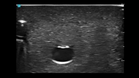
The most superficial structure identified
in the ultrasound image is the sternocleidomastoid muscle.
Deeper to this, the internal jugular vein
will appear as an anechoic elliptical shape
that is compressible with transducer pressure.
In contrast, the anechoic circular artery will be pulsatile.
The thyroid gland lies medial to these structures
and has a light gray echo signature.
In the ultrasound image, the internal jugular vein
will appear superficial to the carotid artery.
Adjust the transducer so the internal jugular vein
is centered within the ultrasound image.
With the needle positioned just superior to the transducer,
slowly insert the needle next to the center line
on the transducer.
The needle will appear as a small bright dot
in the ultrasound image.
When the needle tip appears,
slide the transducer a short distance away,
until the tip is no longer visible.
Once again, advance the needle slowly
until it is visible in the ultrasound image.
Repeat the process to ensure that the needle tip is advanced
under direct ultrasound guidance.
Continue advancing the needle
until the tip is seen to indent
and then puncture the internal jugular vein.
Confirm that the needle tip
lies in the mid portion of the jugular vein
by rotating the probe 90 degrees
to get an in plane longitudinal view of the needle
within the jugular vein.
操作指南:超声引导下内颈静脉中心静脉导管置入术
一段培训视频演示了如何在超声引导下,使用带有静脉检查预设的线阵探头置入内颈静脉导管,操作采用横向入路,患者仰卧且头部转向对侧, 操作者位于床头,探头置于胸锁乳突肌三角下方,定位标记指向9点钟方向。 超声图像显示,胸锁乳突肌为最表浅的解剖结构;内颈静脉表现为位于颈动脉表层的可压缩无回声椭圆形结构,而颈动脉则呈现更接近圆形且具有搏动性;甲状腺位于内侧,回声纹理呈较浅的灰色,有助于区分周围血管解剖结构。 实时针头引导的操作方法是:将针头推进至探头正上方(此时针头在图像中呈现为一个亮点),间歇性地滑动探头以重新定位针尖,并在超声直接显像下反复推进,以确保在整个操作过程中始终追踪到针尖。
相关视频 (1)