
Fujifilm Sonosite Training Hub
All Central Line
Transcript

- [Narrator] This video is for demonstration

and educational purposes only.

Please follow your own institutional guidelines

on sterility and safety.

A linear array transducer with a venous exam type

is used to perform an ultrasound guided insertion

of an internal jugular catheter via a transverse approach.

The patient is in a supine position

with the head slightly turned

towards the contralateral side.

The operator is positioned at the head of the bed.

The transducer is placed transversely just below the apex

of the sternocleidomastoid muscle triangle,

with the orientation marker directed to the operator's left

at a nine o'clock position.

The most superficial structure identified

in the ultrasound image is the sternocleidomastoid muscle.

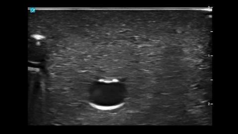
Deeper to this, the internal jugular vein

will appear as an anechoic elliptical shape

that is compressible with transducer pressure.

In contrast, the anechoic circular artery will be pulsatile.

The thyroid gland lies medial to these structures

and has a light gray echo signature.

In the ultrasound image, the internal jugular vein

will appear superficial to the carotid artery.

Adjust the transducer so the internal jugular vein

is centered within the ultrasound image.

With the needle positioned just superior to the transducer,

slowly insert the needle next to the center line

on the transducer.

The needle will appear as a small bright dot

in the ultrasound image.

When the needle tip appears,

slide the transducer a short distance away,

until the tip is no longer visible.

Once again, advance the needle slowly

until it is visible in the ultrasound image.

Repeat the process to ensure that the needle tip is advanced

under direct ultrasound guidance.

Continue advancing the needle

until the tip is seen to indent

and then puncture the internal jugular vein.

Confirm that the needle tip

lies in the mid portion of the jugular vein

by rotating the probe 90 degrees

to get an in plane longitudinal view of the needle

within the jugular vein.

How to: Ultrasound Guided Internal Jugular Central Line

A training video demonstrates ultrasound-guided insertion of an internal jugular venous catheter using a linear array transducer with a venous examination preset, performed in a transverse approach with the patient supine and head turned contralaterally, and the operator positioned at the head of the bed with the probe placed below the sternocleidomastoid triangle and oriented marker at the 9 o’clock position. Ultrasound identification shows the sternocleidomastoid muscle as the most superficial structure, with the internal jugular vein appearing as a compressible anechoic elliptical structure located superficial to the carotid artery, which appears more circular and pulsatile, while the thyroid gland lies medially with a lighter grey echotexture, helping differentiate surrounding vascular anatomy. Real-time needle guidance is performed by advancing the needle just superior to the transducer, visualizing it as a bright dot, intermittently sliding the probe to relocate the tip, and repeatedly advancing under direct ultrasound visualization to maintain tip tracking throughout progression.
Products featured (1)