

Left Ventricular Systolic Function: Eyeball Method
Normal Left Ventricular Function (LVF) Video Clip (0:06)
Here is the same video of a normal LVF.
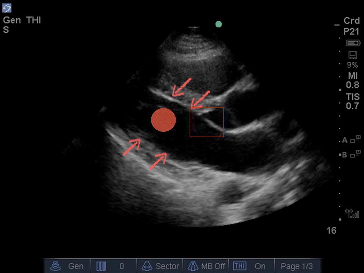
Three Steps – Visually assess for the following:
- Myocardial Thickening
- Endocardial Excursion
- Mitral Valve Leaflet Movement (EPSS)
Using the eyeball method, you can see that is a normal Left Ventricle (LV) in the Parasternal Long Axis (PLAX).
Use these three steps to assess.
- In PLAX view, is there myocardial thickening? To assess for this, look at the muscle (red arrows). Is it getting thicker during the systolic phase of the cardiac cycle?
- Assess for endocardial excursion (red dot). The heart’s walls should generally move towards the center of the chamber. The red dot provides you with a visual goal to look at, and the beginner is encouraged to sometimes just place their finger on the screen, or imagine a similar red dot in place. The two walls pictured in a PLAX indeed move towards the center point.
- In PLAX or apical 4 chamber view, evaluate how closely the anterior Mitral Valve (MV) leaflet comes to the interventricular septum. In this case, during diastole, that anterior MV leaflet comes up and touches the interventricular septum (red box). This is called E-Point Septal Separation (EPSS), a noninvasive echocardiographic evaluation of the LV. It is used for assessing cardiac function. It is well correlated with degrees of systolic function. When used appropriately, EPSS can be an extremely helpful tool in helping assess LVF with the eyeball approach.